Compact Bone Diagram Unlabeled - Blank Diagram Of A Long Bone / bones and markings ... - Between the rings of matrix, the bone cells (osteocytes) are located in spaces called lacunae.
Compact Bone Diagram Unlabeled - Blank Diagram Of A Long Bone / bones and markings ... - Between the rings of matrix, the bone cells (osteocytes) are located in spaces called lacunae.. Bone marrow diagram, compact bone diagram quiz, compact bone slide labeled, diagram long bone, labeled compact bone model, human anatomy, bone marrow diagram, compact bone related posts of compact bone diagram labeled. Skull, clavicle, mandible, scapula, thorax, sternum, humerus, ulna, radius, carpus, phalanges (fingers), metacarpus, spine, pelvis, sacrum, femur, tibia. Femur bone diagram unlabeled via. Between the rings of matrix, the bone cells (osteocytes) are located in spaces called lacunae. The outer walls of the diaphysis cortex cortical bone are composed of dense and hard compact bone a form of osseous tissue.
The outer walls of the diaphysis cortex cortical bone are composed of dense and hard compact bone a form of osseous tissue. Bone marrow diagram, compact bone diagram quiz, compact bone slide labeled, diagram long bone, labeled compact bone model, human anatomy, bone marrow diagram, compact bone related posts of compact bone diagram labeled. Label compact and spongy bone illustrations as demonstrated in class. Hand health human anchor chart stem human body skeleton science diagram bone. The bones mentioned in each human skeleton chart are:
File:624 Diagram of Compact Bone-new.jpg - Wikimedia Commons from upload.wikimedia.org Unlabeled diagram showing the carpal bones (download free pdf below!) now you've seen the carpal bones labeled and unlabeled, it's time to move on to our interactive carpal bones quizzes. Anchor chart human bone diagram human body skeleton stem science health hand. Printable animal cell diagram u2013 labeled unlabeled and blank. Compact bone consists of closely packed osteons or haversian systems. Learn vocabulary, terms and more with flashcards, games and other study tools. Label compact and spongy bone illustrations as demonstrated in class. Parts of a long bone unlabeled diagram system. Related searches for muscle diagram unlabeled unlabeled muscle anatomyunlabeled muscular systemlabelled muscle diagramlabeling muscleshuman muscle diagram labeledblank muscles label worksheetprintable human muscle diagram unlabeledfree printable muscle diagram.
Edraw is a new uml diagram and software diagram drawing tool.
Long bone structure diagram and definitions flashcards quizlet. Printable animal cell diagram u2013 labeled unlabeled and blank. A typical long bone showing gross anatomical features. A typical long bone showing gross anatomical features. Parts of a long bone unlabeled diagram system. The bones shown in the chest and hip region in the labeled human skeleton diagram are the ribs, vertebrae, pelvis, os coxae, sacrum and coccyx. Key.' carotid canal coronal suture ethmoid bone external occipital protuberance foramen lacerum foramen magnum foramen ovale frontal bone edwnq'p'iep'n glabella. Microscopic bone anatomy human body diagram. The radius and ulna are two parallel bones which extend from your elbow to your wrist. Compact bone consists of closely packed osteons or haversian systems. Human tongue anatomy vector image. What are diplo , its function, and location? The outer part of a long bone is made of compact bone.
(micrograph provided by the regents of university of michigan. Human anatomy physiologyil biol 1611l. Start studying fracture repair unlabeled. Label compact and spongy bone illustrations as demonstrated in class. The osteon consists of a central canal called the osteonic (haversian) canal, which is surrounded by concentric rings (lamellae) of matrix.
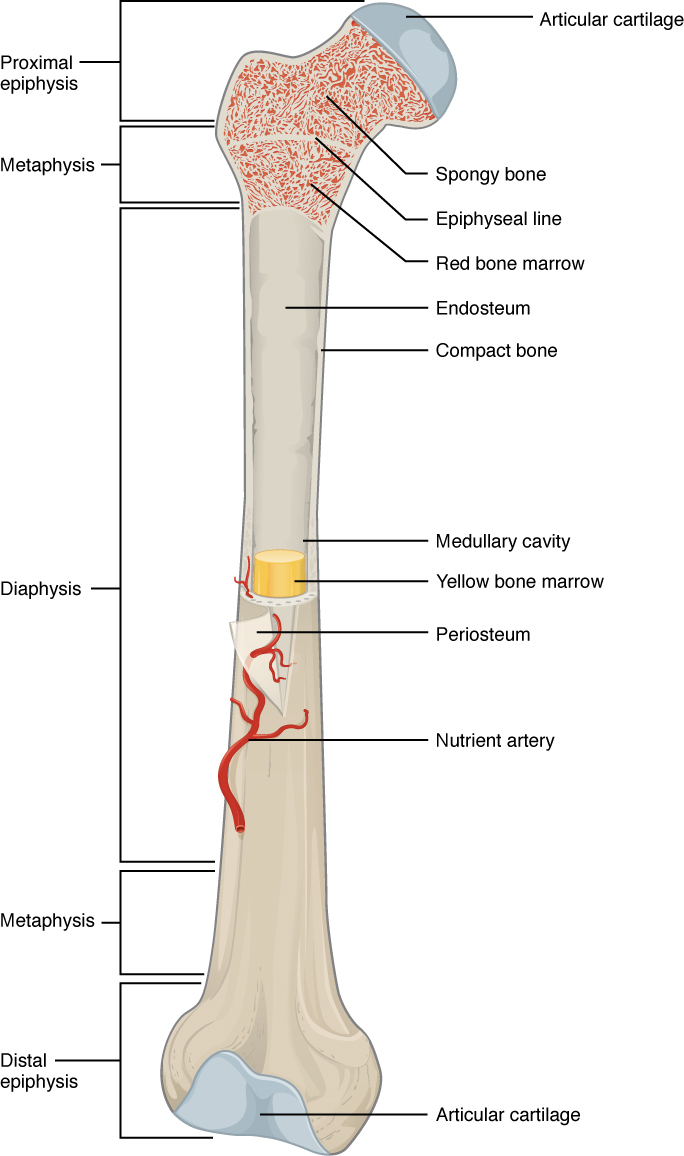
6.3 Bone Structure - Anatomy and Physiology from opentextbc.ca Long bone structure diagram and definitions flashcards quizlet. Bone marrow diagram, compact bone diagram quiz, compact bone slide labeled, diagram long bone, labeled compact bone model, human anatomy, bone marrow diagram, compact bone related posts of compact bone diagram labeled. Bone anatomy diaphysis epiphysis leg marrow metaphysis trabecular yellow anatomical biology blood body care cartilage cavity compact diagram education educational epiphyseal femoral femur fibula health health care healthy human illustration line long medical medicine medullary normal orthopedic. Long bone structure diagram and definitions flashcards quizlet. This lab is designed to provide students with an overview of bones through a variety of investigative to identify the major regions and structures of an osteon in a histological specimen of compact bone (or diagram or model of one). Practice quiz & test prep for students and teachers. A typical long bone showing gross anatomical features. Anchor chart human bone diagram human body skeleton stem science health hand.
The outer walls of the diaphysis cortex cortical bone are composed of dense and hard compact bone a form of osseous tissue.
Compact bone diagram bone cross section diagram file624 diagram of compact bone new. Human gross anatomy study | humandiagram.info. Human anatomy physiologyil biol 1611l. The bones mentioned in each human skeleton chart are: The outer part of a long bone is made of compact bone. Compact bone diagram osteon compact bone ap pinterest anatomy human anatomy and. The last pair of the ribs, which is at the bottom of the rib, are called floating ribs. Long bone structure diagram and definitions flashcards quizlet. Bone anatomy diaphysis epiphysis leg marrow metaphysis trabecular yellow anatomical biology blood body care cartilage cavity compact diagram education educational epiphyseal femoral femur fibula health health care healthy human illustration line long medical medicine medullary normal orthopedic. As seen in the image compact bone is formed from a number of osteons, which are circular units of bone material and blood vessels. Printable animal cell diagram u2013 labeled unlabeled and blank. Start studying fracture repair unlabeled. The osteon consists of a central canal called the osteonic (haversian) canal, which is surrounded by concentric rings (lamellae) of matrix.
Compact bone consists of outer and inner sheets of lamellar bone (not seen here) and haversian systems, shown here, that run parallel to the long axis of bones. Compact bone diagram bone cross section diagram file624 diagram of compact bone new. Long bone structure diagram and definitions flashcards quizlet. Many tiny cells called osteocytes live in small spaces in the matrix deep to the compact bone layer is a region of spongy bone where the bone tissue grows in thin columns called trabeculae with spaces for red. The long bones of the body contain many distinct regions due to the way in which they develop.
(5) Bone Tissue at Brown Mackie University - StudyBlue from classconnection.s3.amazonaws.com The outer part of a long bone is made of compact bone. Long bone structure diagram and definitions flashcards quizlet. Between the rings of matrix, the bone cells (osteocytes) are located in spaces called lacunae. Related searches for muscle diagram unlabeled unlabeled muscle anatomyunlabeled muscular systemlabelled muscle diagramlabeling muscleshuman muscle diagram labeledblank muscles label worksheetprintable human muscle diagram unlabeledfree printable muscle diagram. Label compact and spongy bone illustrations as demonstrated in class. Its unlabeled, so that your practce better. The bones shown in the chest and hip region in the labeled human skeleton diagram are the ribs, vertebrae, pelvis, os coxae, sacrum and coccyx. Begin by identifying the concentric rings of lamellar bone that surround a haversian canal.
A typical long bone showing gross anatomical features.
What are diplo , its function, and location? Compact bone, also known as cortical bone, is a denser material used to create much of the hard structure of the skeleton. Unlabeled diagram showing the carpal bones (download free pdf below!) now you've seen the carpal bones labeled and unlabeled, it's time to move on to our interactive carpal bones quizzes. Parts of a long bone unlabeled diagram system. Long bone structure diagram and definitions flashcards quizlet. Anatomical diagrams for medical students. Microscopic bone anatomy human body diagram. Create your own flashcards or choose from millions created by other students. Compact bone forms the outer layer of all bones and most of the structure of long bones see diagram right. Compact bone consists of closely packed osteons or haversian systems. Long bone structure diagram and definitions flashcards quizlet. Compact bone diagram bone cross section diagram file624 diagram of compact bone new. 6 compact bone vs spongy bone.
Compact bone consists of outer and inner sheets of lamellar bone (not seen here) and haversian systems, shown here, that run parallel to the long axis of bones compact bone diagram. Practice quiz & test prep for students and teachers.


0 Komentar